

PIGMENTATION IN BLACK & MATTE SKIN
Pigment synthesis:
Melanogenesis is the scientific term for skin pigmentation. Its evolution takes place at the level of the epidermis, the outermost layer of the skin; in particular dendritic cells: melanocytes. The latter interact with nearby keratinocytes to homogeneously diffuse the synthesized melanin. The process of melanogenesis is carried out within specific organelles called melanosomes, organelles intrinsic to melanocytes. They are therefore the support for the synthesis of different pigments (melanins), responsible for our specific skin color (phototypes).
Skin physiology
The skin accounts for about 15% of body weight in adults and has an area between 1.5 and 2m². It is the largest organ in the human body. Its main role is its function as a physical barrier that it represents against mechanical, chemical, physical or microbiological aggressions; but it also has sensory properties (touch, pressure...), thermoregulatory (modulation of blood flow), metabolic properties (vitamin D synthesis, energy reserve...), immune, and a unique defense system against UV rays: pigmentation.
It is composed of four different strata (Fig. 1):
- The EPIDERMIS is a multi-layered tissue without a nerve and vascular system. It has several cell types: keratinocytes up to 95%; melanocytes (seat of pigmentation at the basal layer); Langerhans cells (immune role).
- THE DERMO-EPIDERMAL JUNCTION: is acellular, non-vascularized and is the support of the anchor structures between the dermis and the epidermis. It has a role in maintaining the proliferative potential of basal keratinocytes.
- THE DERMIS is the supporting tissue of the epidermis, but also the sensory region of the skin (vascular and nervous networks, mechanoreceptors ...). It contains epidermal appendages such as the sebaceous glands responsible for the secretion of sebum which allows the maintenance of the suppleness and impermeability of the skin and limits the insensitive loss of water (PIE).
- HYPODERMIS is the subcutaneous fatty tissue.
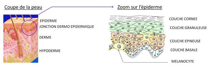
Figure 1: Presentation of a skin cut and zoom on the epidermis
The synthesis of melanin is carried out in specific cells, melanocytes, at the level of the basal layer of the epidermis. These dendritic cells (= cell extensions), are the support of particular organelles called melanosomes. The latter are small vacuoles allowing the transport and release of the melanin formed, from the melanocyte to the surrounding keratinocytes, and thus allow the dispersion of pigmentation. The peculiarity of black skin lies in the fact that this melanin is not degraded during its passage through the upper layers of the epidermis. It thus reaches the stratum corneum intactly, with the great difference of Caucasian skin (Fig. 2 ).
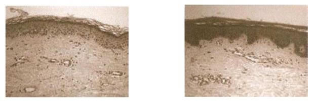
Figure 2: Microscopic views of a Caucasian skin section (left) and a black skin cut (right)
The different phototypes
Each individual has a skin color of their own. Skin pigmentation, in different people, is none other than a mosaic of colors that is declined to infinity. Skin color is an intrinsic characteristic of a given individual. There are six main classes and these are called phototypes (Table 1 & Fig. 3).
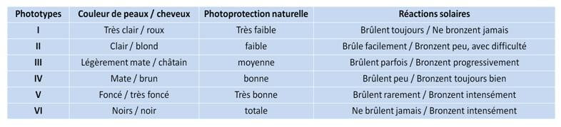
Table 1: Presentation of the six phototypes and their characteristics
These different phototypes are determined genetically. This implies the definitive and personal nature of the skin color.

Figure 3: Photos of the different phototypes
It may seem strange, but melanocytes, specific cells of melanogenesis, are in identical numbers in all phototypes. The difference in skin color therefore comes from another parameter. This implies a variation in the functioning of the process of melanogenesis, which results in a more or less important production of melanin in each individual.
The pigmentation process: melanogenesis
The melanogenesis responsible for pigmentation is a response to external stimulation: ultraviolet (UVs). This is called induced pigmentation, more commonly known as "tanning". This differs from the so-called basal constitutive pigmentation, specific to an individual. Through the synthesis of melanin, the skin protects itself from physico-chemical aggressions, thus reducing the penetration of UV rays (Fig. 4). This mechanism is also called natural photo-protection.
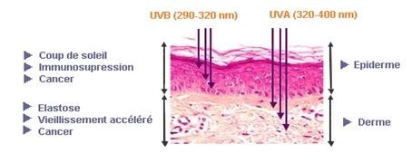
Figure 4: Diagram of skin penetration by UV rays
As we have seen previously, the site of melanogenesis is melanosome, the melanocyte-specific organelle. Melanosome is the medium in which maturation and deposition of melanin occur. The melanin grains are carried by membrane proteins to the ends of each melanocyte dendrite, to transfer their contents to the surrounding keratinocytes in order to dissipate the color homogeneously. We also saw that the number of melanocytes was not the cause of interethnic variations since it is the same regardless of the skin color of the individual. It is therefore essential to detail the mechanism of melanogenesis in order to understand its functioning, its regulation and thus be able to act as best as possible to regulate it.
The process of melanogenesis is a complex biochemical mechanism that involves many enzymes, such as tyrosinase, TYRP-1 and dopachrome tautomerase (TYRP-2). (Fig.5). All of them play a fundamental role during the synthesis of melanin and allow the regulation of pigmentation.
Tyrosinase (or DHI oxidase):
- melanogenesis limiting enzyme
- catalyzes the first two reactions of the process
- also catalyzes the formation of the indole-5,6-quinone complex from DHI
- is necessary for the transformation of DHICA into an indole-5,6-quinone-2-carboxylic acid complex by TYRP-1
TYRP-1 or DHICA-oxidase:
- Catalyzes the oxidation reaction of DHICA into an indole-5,6-quinone-2-carboxylic acid complex
- Works synergistically with tyrosinase
- Stabilizes Tyrosinase
Dopachrome tautomerase or TYRP-2:
- Catalyzes the formation of DHICA from Dopachrome
Melanocytes are able to synthesize two pigments of a non-similar nature: pheomelanins (yellow / red carcinogenic pigments) and eumelanins (brown pigments, photoprotective blacks), from the same precursor: L-Tyrosine. This variation is due to the melanin synthesis pathway which involves very specific and distinct enzymes. Each individual presents the two pigments but in different proportions. It can therefore be said that the variability of pigments depends on the genetic equipment that codes for these enzymes. Caucasian skin strongly expresses the gene that codes for tyrosinase, unlike black and matte skin, which largely express the genes encoding the other two enzymes: dopachrome tautomerase (TYRP-2) and DHICA oxidase (TYRP-1).
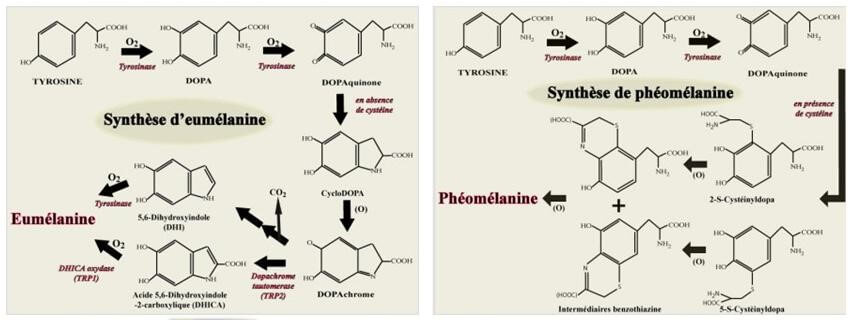
Figure 5: Diagram of the melanogenesis process
Conclusion
Melanogenesis is a complicated process to modify, as it is genetically programmed, but it can be modulated. In black and matte skin, this mechanism gets carried away very quickly and can be the cause of spotted and non-homogeneous skin. In this case we speak of hyperpigmentation, the most frequent cause of consultations with dermatologists. The MEL'OYA® range has been developed specifically to provide an effective solution to the problems of hyperpigmentation and unification of the complexion.